An international team of scientists, with significant participation from researchers at the IBIMA institute in Málaga, has made a relevant advance in the study of glioblastoma, a type of brain tumor known for its aggressiveness and poor prognosis. The research has focused on the use of cutting-edge materials and advanced techniques to analyze three-dimensional (3D) models of this tumor.
The study, jointly led by Tanja Dučić, a scientist at ALBA Synchrotron and professor at the University of Belgrade, and Elena González, head of a research group at IBIMA Plataforma BIONAND and professor of Cell Biology at the University of Málaga, employed a nanocomposite activated and analyzed with synchrotron light. The results, reported by the Junta de Andalucía, show a significant response in the 3D glioblastoma models.
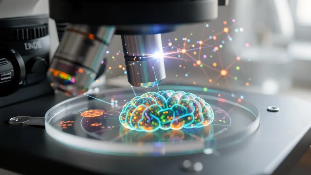
For the research, 3D spheroids of patient-derived glioblastoma cells were used, comparing them with astrocyte spheroids as a non-tumoral control. The objective was to observe the response of these complex biological systems to a nanocomposite based on 'carbon dots' (nanomaterials) and the drug riluzole, evaluating the resulting molecular modifications.
The resistance of glioblastoma to conventional treatments and its molecular heterogeneity have driven the need to develop more precise experimental models. Three-dimensional spheroids, which better mimic the architecture and cellular communication of a real tumor, have been key to this study, offering a more realistic experimental environment for analyzing tumor response to new therapeutic approaches.
The results from the nanocomposite, which combines 'carbon dots' and riluzole, have revealed detectable molecular modifications in the glioblastoma spheroids, particularly in spectral regions associated with nucleic acids, lipids, and proteins. These alterations were more pronounced in the tumoral models than in the astrocyte models, demonstrating a differential response.
Infrared microspectroscopy, using high-intensity and precision synchrotron radiation, has acted as a molecular 'super-microscope'. This non-invasive technique has allowed for the detection of subtle biochemical changes in proteins, lipids, and nucleic acids without the need for chemical markers, mapping the treatment effects with unprecedented resolution.
The analysis has revealed alterations in cancer cell DNA, signs of oxidative stress, and modifications in protein folding, compromising glioblastoma viability. A promising finding is the selectivity of the treatment, identifying clearly differentiated molecular responses between glioblastoma and astrocyte spheroids, which provides crucial information about cellular sensitivity.